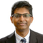
DR ASHNIL KUMAR
BEng(Software)/BArts Sydney, PhD Sydney
Postdoctoral Research Fellow
University of Sydney
Dr Ashnil Kumar received his PhD from the University of Sydney in 2013. He is currently a postdoctoral research fellow in the School of Information Technologies conducting research into medical image analysis algorithms and their application within clinical workflows. He works closely with clinicians from a number of hospitals in Sydney, including the Royal Prince Alfred Hospital, Westmead Hospital and Nepean Hospital. His research focuses on deriving analysis algorithms that align with the semantic understanding of clinical experts across a variety of different imaging data. His algorithms have been used for cancer analysis in PET-CT, fetal development monitoring in ultrasound, skin cancer prediction in dermoscopic imaging, and event detection in cell videos.
Research Highlight 1
I developed a technique to analyse PET-CT images to construct a graph model that represented the visual properties of anatomical structures and tumours, as well as their spatial relationships.
This work resulted in a graph-based image retrieval algorithm that measured image similarity based upon criteria such as the spatial proximity of tumours in relation to specific organs, which can be an important clinical biomarker.
Research Highlight 2
I developed several techniques to enable visual abstraction of image retrieval information and to allow exploration of complex image feature spaces.
RESULT
This work resulted in user interface designs visual analytics tools that allowed the human users to better understand the characteristics that made different images similar, thereby acting to bridge the semantic gap, and opening the door for use of retrieval within computerised decision support systems.
Research Highlight 3
I developed an algorithm that used an image retrieval framework to analyse and automatically annotate lesion-bearing liver CT images.
RESULT
This algorithm won the international ImageCLEF Liver CT Annotation Challenge in 2014 and its accuracy was unbeaten in 2015.
Research Highlight 4
I have developed algorithms to identify and measure body structures in fetal ultrasound images.
RESULT
These algorithms were used to identify the fetal brain and to measure the thalamic diameter, a possible biomarker for neurodevelopment. The measurement accuracy was consistent with a sonographer with 20 years of experience.
Research Highlight 5
I am part of a team that is adapting deep neural networks to enable the analysis of medical images and videos.
RESULT
We have developed deep neural networks to identify different types of medical images, to detect melanoma in dermoscopic images, to identify cell divisions in video data, and to learn image properties with limited training labels.

